Written by Dr. Lance Kisby
Introduction
The term Molar Incisor Hypominerlaization (MIH) was first introduced in 2001 to describe ‘hypomineralisation of systemic origin, presenting as demarcated, qualitative defects of enamel of one to four first permanent molars (FPMs) frequently associated with affected incisors.’ In 2003, it was further defined as “a developmental, qualitative enamel defect caused by reduced mineralization and inorganic enamel components which leads to enamel discoloration and fractures of the affected teeth.”2 Initially, the condition was described as affecting the first permanent molars (FPMs) and incisors but more recently it has been noted that these defects could affect any primary or permanent tooth.3 Weeheijam showed they can also occur on second primary molars, permanent molars, and the cusp tips of permanent canines.2
In its mildest condition, the enamel can appear white-yellow and in its more severe condition, the enamel can be brown-orange. The discoloration is easy to differentiate from other enamel defects in that the effected areas are asymmetric with irregular borders.4
Treating pediatric dental patients with MIH pose several challenges.5-8 Severe post eruptive breakdown is common on stress bearing teeth. When the hypomineralization is on the occlusal of primary and permanent molars the result can be post eruptive breakdown which causes these teeth to be sensitive to cold. Consequently, there is, very often, an inability to achieve adequate local anesthesia and it is thought to be, possibly, related to chronic pulpal inflammation. In pediatric patients, behavior guidance problems result due to dental fear and anxiety resulting from the pain experienced from previous multiple treatment appointments.
Treating these teeth is also challenging. In children, MIH incisors create esthetic concerns which has been shown to have compromised functioning, well-being, quality of life,9 anxiety, depression, and other mood states.10
Treatment options
There are many treatment options available in young patients for anterior MIH incisor teeth. The best treatment is a conservative approach as these immature anterior teeth have large and sensitive pulps.11 Composites require removing the effected area, a situation where local anesthesia may not work, require long term observation and maintenance due to discoloration, wear, and marginal fractures.12
Porcelain veneers are indicated for patients over age 18 years after the gingival margin has matured and is best used when other more conservative techniques have failed.12-13
Resin infiltration has many benefits. The refractive index of enamel is 1.62 and the refractive index of resin infiltration is 1.52. This technique improves the translucency and thus improves esthetics.14-15
This article will present a conservative treatment option of Icon Resin Infiltration of anterior permanent teeth with MIH in children. *Please note that while MIH is not an approved Icon indication, the resin infiltration technique has been shown to be effective in several cases for correcting esthetic issues of MIH lesions on anterior teeth.
Case #1
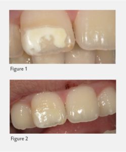
Figure 1 shows a well demarcated MIH buccal white lesion on tooth #8. The patient, an 8–year–old girl, presented with her mother concerned about the white spot on #8. The patient related how friends were teasing her at school, calling her names, and the mother related that the patient has no friends.
Icon resin infiltration system consists of an etch of 15% HCI, Icon Dry which is 99% ethanol, and a methacrylate based resin.
The technique used is as follows: the tooth is isolated with a rubber dam. With a carbide finishing bur, remove the thin surface layer of the lesion in order for the infiltrate resin to gain access to the lesion body. Next, Icon Etch was placed by massaging the Etch over the surface for a 2-minute period. The instructions call for inspecting the tooth to get a preview of the final result by rinsing off the etch, placing Icon Dry and the whitish-opaque area should diminish. If not, the etch and preview step can be repeated up to 2 more times. In this case, after three times of etching, rinsing, and previewing, there was no change in color. I decided to repeat both the etch and preview steps.
At the last preview step, there was only a very slight change in color. It was decided to stop etching for fear of removing too much enamel.
As indicated in the Icon instructions, the tooth was dried with an oil free air syringe. Icon Dry was placed onto the lesion and allowed to set for 30 seconds. For best treatment results, it is necessary to dry the lesion again with an oil free air syringe. Now that the lesion is completely desiccated, the tooth is ready to absorb the infiltrating resin. The lcon lnfiltrant cannot be applied under direct operatory light because the material will set prematurely. After all lights in the room were shut off, an ample amount of lcon lnfiltrant was placed onto the etched and dried surface by continuously turning the shaft of the syringe and massaging the resin material into the prepared lesion with the applicator to keep the surface wet. I determined this lesion to be deeper and larger than most MIH defects. The instructions indicate the esthetic result of the resin can be improved by extending the penetration of the resin for up to 6 minutes, which was done in this case, instead of the usual 3 minutes. The lcon lnfiltrant was light cured for 40 seconds. The resin was applied a second time, allowed to penetrate for 1 minute, and then light cured for another 40 seconds. The surface was gently smoothed with polishing cups. Figure 2 shows the final result.
At the one-month follow-up, the mother related that the patient is now smiling more, happier, making new friends, and is now getting invited to sleep-overs. I noticed the patient was smiling more and seemed happier than the first time I had seen her.
Case #2
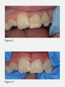
Figure 3 shows an MIH lesion on the buccal of #9. This patient was a 9–year–old boy. The technique was performed as in Case #1 except the lesion was etched with the Icon Etch 5 times. After each etch, the Icon Dry was used to preview the result. There was still no difference in color of the lesion after the fifth etch. I decided to etch fewer times to decrease the amount of enamel loss.
Figure 4 shows the final result where the lesion color matches the enamel of the rest of the tooth.
Discussion
MIH effected enamel is characterized by a reduction in mineral quality as well as an increased porosity.16 Molars with MIH have 5 to 10 times more treatment than molars with no MIH.17
The most commonly encountered problems in MIH affected anterior teeth are thermal hypersensitivity, discoloration, and enamel break down.18 Young patients frequently comment on esthetic concerns regarding anterior teeth which can lead to psychosocial issues.
From the above post treatment images, the Icon Resin Infiltration technique creates excellent and pleasing esthetic results on MIH anterior teeth. It is a conservative alternative to complete removal of the lesion on anterior teeth.
The etching instructions for Icon Resin Infiltration are indicated for etching white spots in enamel commonly seen after orthodontic bands and brackets have been removed. MIH enamel white lesions on anterior teeth are the result of a different process than white spots from caries on the same teeth. This would account for why 2–to–3 etching cycles are appropriate for enamel caries while additional etching cycles were used to change the MIH lesion color. More research is needed for the optimal time for etching MIH anterior teeth. Less etching time would preserve more tooth structure and take less time, an important consideration when doing this procedure on pediatric patients.
While MIH is not an approved indication for Icon, the resin infiltration technique has been shown to be effective in several cases for correcting esthetic issues of MIH lesions on anterior teeth without local anesthesia; an important behavior guidance consideration when dealing with pediatric patients who may have been traumatized by previous restorative attempts.
- References
Weerheijm K L, Jalevik B, Alaluusua S . Molar-incisor hypomineralisation. Caries Res 2001;5: 390-391. - Weerheijm K L, Duggal M, Mejare I et al. Judgement criteria for molar incisor hypomineralisation (MIH) in epidemiologic studies: a summary of the European meeting on MIH held in Athens, 2003. Eur J Paediatr Dent 2003; 4: 110-113.
- Steffen R, Van Waes H . Therapy of MolarlncisorHypomineralisation under difficult circumstances. A concept for therapy. Quintessenz 2011; 62: 1613-162.
- Allazzam SM, ALAKI SM, Meligy OAS. International J Denti. 2014. Doi. Org /10.1155/2014/234508.
- Kalkani M, Balmer R C, Homer R M, Day P F, Dug gal M S. Molar incisor hypomineralisation: experience and perceived challenges among dentists specialising in paediatric dentistry anda group of general dental practitioners in the UK. Eur Arch Paediatr Dent 2016; 17: 81-88.
- Ghanim A, Silva M J, Elfrink M EC et al. Molar incisor hypomineralisation (MIH) training manual for clinical field surveys and practice. Eur Arch Paediatr Dent 2017; 18: 225-242.
- AI-Batayneh O B, Jbarat RA, AI-Khateeb S N. Effect of application sequence of fluoride and CPP-ACP on remineralization of white spot lesions in primary teeth: An in-vitro study. Arch Oral Biol 2017; 83: 236-240.
- Kalkani M, Balmer RC, Homer RM, Day PF, Duggal MS. Molar incisor hypomineralisation: experience and perceived challenges among dentists specializing in paediatric dentistry anda group of general dental practitioners in the UK. Eur Arch Paediatr Dent. 2016;17:81-88.
- Almauallem 2., Busuttil-Naudai A. Molar incisal hypomineralization (MH)- an overview. Brit Dent J. Oct. 18, 2018 225(7):601-609.
- Settineri, S., Rizzo, A., Liotta, M., & Mento, C. (2017). Clinical Psychology of Oral Health: The Link Between Teeth and Emotions. SAGE Open, 7(3). https://doi.org/10.1177/ 2158244017728319
- Ghanim A, Silva M J, Elfrink M E C et al. Molar incisor hypomineralisation (MIH) training manual for clinical field surveys and practice. Eur Arch Paediatr Dent 2017; 18: 225-242.
- Lygidakis NA. Treatment modalities in children with teeth affected by molar-incisor enamel hypomineralisation (MIH): A systematic review. Eur Arch Paediatr Dent 2010; 11: 65-74.
- Wray A, Welbury R. Treatment of intrinsic discoloration in permanent anterior teeth in children and adolescents. 2004. Available at https://www.rcseng. ac.uk/-/med ia/files/rcs/fds/publications /discolor. pdf.
- Comisi J C. Provisional materials: advances lead to extensive options for clinicians. Compend Contin Educ Dent 2015; 36: 54–59.
- Attal JP, Atlan A, Denis M, Vennat E, Tirlet G. White spots on enamel: treatment protocol by superficial or deep infiltration (part 2). Int Orthod 2014; 12: 1-31.
- Elhennawy, K. et al. Structural, mechanical and chemical evaluation of molar-incisor hypomineralization-affected enamel: A systematic review. Arch. Oral. Biol. 83, 272-281.
- Jalevik B, Klingberg G. Treatment outcomes and dental anxiety in 18-year-olds with MIH, comparisons with healthy controls-a longitudinal study. Int J Paediatr Dent. 2012;22(2):85-91.
- Souza, Juliana. Aesthetic management of molar-incisor hypomineralization. Revista Sul-brasileira de Odontologia. 2014; 11: 204
