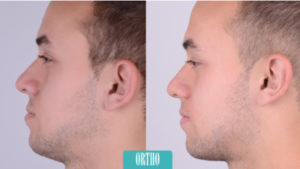
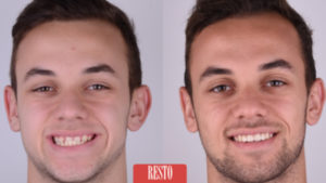
Written by Igor Ristić, DDS, Center for Dental Aesthetics and Implantology, Belgrade
Treatment options for solving aesthetic problems caused by missing lateral incisors, previously involved standard solutions, including often less conservative approaches such as implant placement or preparation of neighboring teeth for FPR. Maryland type bonded restorations often showed lower survival rate due to intrusion and also caused debonding. Additionally, implant placement in young patients is contraindicated due to following facial growth while exposing patients to a high biological cost during crown preparation, which is no longer acceptable from both ethical and professional points of view.
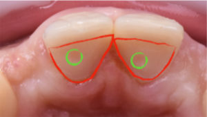
Solving problems with RBFPD (Resin Bonded Fixed Partial Denture) is not new. It began with PFM structure that was cemented and then bonded. It then evolved to Zirconia framework, but with the introduction of MDP monomer and its availability in resin cements. The suggested treatment option showed very promising survival rate and viable therapeutic option for such demanding cases.
It challenges the clinician in occlusion diagnostics, preparation design, Zirconia surface pretreatment, bonding protocol, and also the whole team with lab technician in creating perfect natural aesthetic from ceramic material. Described treatment can be successful for patients with cleft palate aesthetic problems (often retained on canine) or with unilaterally missing lateral incisor.
Treatment phases in brief
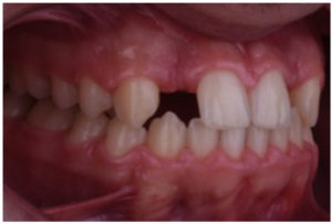
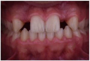
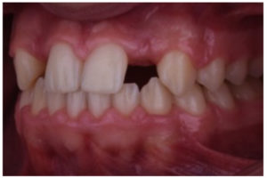
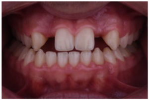
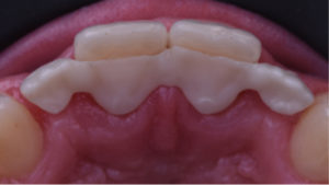
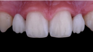
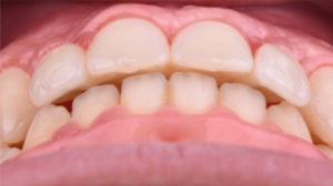
1. After orthodontic treatment, the patient was referred for restorative treatment. Initial intra end extraoral, static and dynamic occlusion check images below. (Figures 1-4)
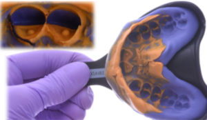
2. Preparation was conducted in enamel, creating a chamber and pinhole to place the Zirconia framework (5881 bur and Soniflex tip SF979, Komet/Brasseler). It can usually be placed out of the occlusal contact area. (Figure 5) The pinhole is essential for repeated and precise placement during trials, lab phases and bonding positioning. After tissue management with two cords technique (Ultrapak 000 and 1, Ultradent), impressions are made with A-silicone in single step technique (Honigum Heavy Body and Honigum Light Body, DMG), and plaster
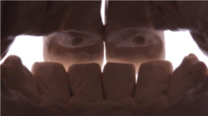
models are made. (Figure 6) This preparation design is very convenient for intraoral scaring procedure. (Figure 7)
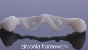
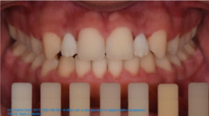
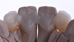
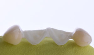
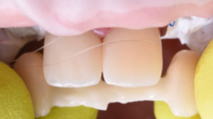
3. Framework is made of LT high strength Zirconia (ZirCAD, Ivoclar Vivadent), intraoral trial is essential for fit check and its position related to natural hard and soft tissues. (Figure 8, 9, 10) Adequate shade selection is essential for straight forward workflow. Use of polarized filters in dental photography give possibility to determine value of the color. (Figure 11)
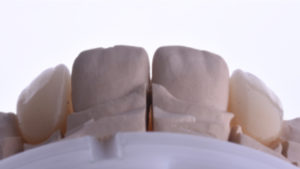
4. Final form with veneering with Emax Ceram ceramics (Ivoclar Vivadent). (Figure 12) Ovate pontic form in cervical areas of lateral incisors would influence final soft tissue contour. In this case soft tissue tunnel graft was applied to mask missing bone volume in area 12 and 22. (Figures 13, 14)
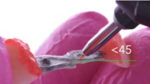
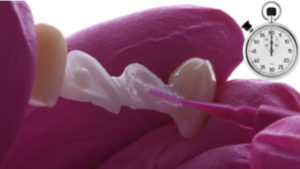
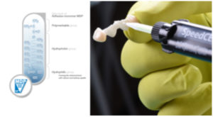
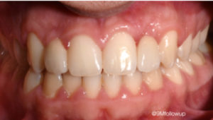
5. After the protection of glared ceramics (Pafern Resin, GC), the bonding procedure requires sandblasting procedure (20s exposure, 0.2 Mpa, 110 microns grid), cleaning, coating with MDP monomer (Monobond Plus, Ivoclar Vivadent), 37% phosphoric acid enamel etching rinsing and dry step, and the use of self etch resin cement with MDP monomer (Speedcem Plus, Ivoclar Vivadent). The bonded area is light cured from both aspects 40 seconds with air cooling. Proper rubber dam isolation was not applied due to synchronised soft tissue grafting procedure. Teflon tapes and retraction cords (Ultrapack, Ultradent) were used. (Figures 15-17) Intraoral photos made on 8 weeks follow up. (Figures 18-20)
6. The concept of RBFPD is often used as addition to orthodontic therapy, with possibilities of added soft tissue grafting or some additional non-invasive therapeutic options could be recommended in restoring missing LI. Implant treatment could always be an option in later stages of life. This patient expressed his full compliance during the treatment phase and satisfaction with the final results.
Literature
1. Heij DG1, Opdebeeck H, van Steenberghe D, Kokich VG, Belser U Facial development, continuous tooth eruption, and mesial drift as compromising factors for implant placement. Int J Oral Maxillofac Implants. 2006 Nov-Dec;21(6):867-78
2. Kern M, Passia N, Sasse M, Yazigi C Ten-year outcome of Zirconia ceramic cantilever resin-bonded fixed dental prostheses and the influence of the reasons for missing incisors J Dent. 2017 Oct;65:51-55
3. Sekhri S, MiOal S, Garg S,Tensile Bond Strength of Self Adhesive Resin Cement After Various Surface Treatment of Enamel. J Clin Diagn Res. 2016 Jan;10
4. Cardenas AM, Siqueira F, Hass V, Malaquias P, Gutierrez MF, Reis A, Perdigão J, Effect of MDP-containing Silane and Adhesive Used Alone or in Combination on the Long-term Bond Strength and Chemical Interaction with Zirconia Ceramics. J Adhes Dent. 2017;19(3):203-212.
5. Su N, Yue L, Liao Y, Liu W, Zhang H, Li X, Wang H, Shen J The effect of various sandblasting conditions on surface changes of dental Zirconia and shear bond strength between Zirconia core and indirect composite resin. J Adv Prosthodont. 2015 Jun;7(3):214-23 Figure 15 Figure 16 Figure 17 Figure 18 Figure 18 Figure 20
6. EinarsdoZr ER, Lang NP, Aspelund T, Pjetursson BE A multicenter randomized, controlled clinical trial comparing the use of displacement cords, an aluminum chloride paste, and a combination of paste and cords for tissue displacement. J Prosthet Dent. 2018 Jan;119(1):82-88.
7. Kim TH1, Cascione D, Knezevic A. Simulated tissue using a unique pontic design: a clinical report. J Prosthet Dent. 2009 Oct;102(4):205-10
8. Sasse, M., Kern, M. CAD/CAM single retainer Zirconia-ceramic resin-bonded fixed dental prostheses: clinical outcome after 5 years. International journal of computerized dentistry Volume 16, Issue 2, 2013, Pages 109-118
9. Antonarakis GS, Prevezanos P, Gavric J, Christou P. Agenesis of maxillary lateral incisor and tooth replacement Cost-Effectiveness of different treatment alternatives. Int J Prosthodont. 2014;27:257–63.
10. Araujo EA, Oliveira DD, Araujo MT. Diagnostic protocol in cases of congenitally missing maxillary lateral incisors. World J Orthod.2006;7:376-88
11. Ghuman T, Donovan T. Effect of MDP containing self-adhesive cements bond strength to Zirconia. J Dent Res. 2015;94(Spec Iss A):3659
12. Magnuszewski T, et al. Moisture effect on shear bond strength of self-adhesive resin cements. J Dent Res. 2016;95(Spec Iss A):587.
